

Embarking on the journey of pregnancy through In Vitro Fertilisation (IVF) is a momentous occasion filled with hope, anticipation, and careful planning. After navigating fertility treatments, every milestone during your pregnancy brings both excitement and a desire to ensure everything is progressing smoothly.
As part of your routine prenatal care, your obstetrician may recommend a specialized imaging procedure known as a fetal echocardiogram (or fetal echo for IVF pregnancy). If you have been advised to get this scan, it is completely natural to wonder why it is necessary, how it differs from routine scans, and what to expect during the procedure.
At RNK Diagnostics, we aim to empower expecting parents with clear, compassionate, and accurate medical information so you feel supported at every step of your pregnancy journey.
What Is a Fetal Echocardiogram?
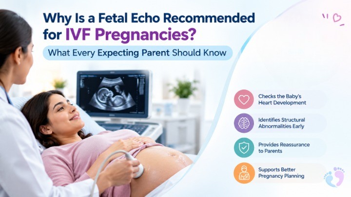
A fetal echocardiogram is a unique form of ultrasound for obtaining images of the baby’s heart before birth. In contrast to most other types of prenatal ultrasound tests that provide information about the baby’s growth and movement, a fetal echocardiogram provides a detailed view of the chambers of the baby’s heart, as well as its valves and various arteries.
Performed typically between 18 and 24 weeks of gestation typically, this non-invasive test uses safe sound wave technology—free of any radiation—to observe cardiac development in real time.
Why Is a Fetal Echo Needed in IVF Pregnancies?
If you conceived through IVF, then you might question why another scan is needed when the early ultrasound results are good. Here are the main reasons why doctors recommend a fetal echocardiogram after IVF:
1. Slight Statistical Variance in Cardiac Development
Clinical studies in the field of reproductive medicine show a small statistical increase in congenital heart defects in infants who have been conceived using assisted reproductive technology (ART) such as IVF or ICSI. The overall absolute risk is quite low and continued monitoring provides peace of mind.
2. Comprehensive Evaluation of Early Organ Formation
During the delicate early stages of embryology in IVF cycles, multiple factors—such as underlying parental conditions, hormonal stimulation, or culture environments—can influence subtle development. A targeted scan allows specialists to verify that heart structures have formed seamlessly.
3. Early Detection and Early Action
Detecting potential cardiac variations during pregnancy gives parents and delivery teams the advantage of advance planning. If a minor condition is identified, medical care can be tailored for delivery at a facility equipped with specialized neonatal support, ensuring optimal safety for the baby.
Fetal Echo vs. Standard Anomaly Scans: What’s the Difference?
During your second trimester, you will likely undergo a routine mid-trimester anomaly scan, often enhanced by advanced imaging techniques like a 4D ultrasound in Dwarka to visualize detailed facial features, limb structure, and soft tissue.
While conventional two-dimensional (2D) and three-dimensional (3D) imaging technology is able to produce high-quality images of most of the anatomical structures, the heart is a complicated organ that works quite fast. Echocardiography during the IVF pregnancy means that high-quality probes are being used along with Doppler flow mapping and cardiac protocol, which enables tracking the blood flow inside the heart chambers.
Is a Fetal Echo Safe for IVF Babies?
Certainly. Fetal echocardiography is absolutely safe both for mother and fetus. This method uses conventional high-frequency sound waves, similar to normal obstetric ultrasonography. This procedure does not cause any pain; it does not require needle puncture, and there is no need for any special preparation or diet.
What Happens During the Procedure?
When you schedule your scan:
- Comfortable Setup: You will relax comfortably on an examination couch while a specialized sonographer or fetal cardiologist applies warm gel to your abdomen.
- Detailed Imaging: The transducer probe gently glides across your stomach to capture precise views of your baby’s heart anatomy and circulation patterns.
- Duration: The scan typically takes between 30 to 45 minutes, depending on the baby’s position.
- Immediate Insights: In most cases, the specialist can discuss initial findings with you right after the appointment.
Prioritizing Fetal Health and Peace of Mind
A specialist heart scan for your baby is something that you should not worry about. In today’s times, medical specialists are more inclined towards suggesting this test just as a precautionary method to keep your baby safe and sound. Knowing what to expect from the visit will help you enjoy meeting your precious little angel without any worries about her heart health.
Choosing a trusted diagnostic center equipped with high-resolution imaging technology and experienced fetal medicine specialists makes all the difference in your care experience. If you are seeking reliable prenatal care, advanced diagnostic services, and supportive patient management, scheduling a dedicated Fetal echo in Dwarka at RNK Diagnostics ensures that your pregnancy journey is guided by technical precision, clinical accuracy, and empathetic expertise every step of the way.
Key Takeaway for Parents
IVF makes you a very dedicated and loving woman, among many other things. Getting a referral for your baby’s fetus to undergo a fetal echocardiogram does not necessarily mean that anything is wrong. It is simply a precaution that helps confirm whether everything is okay or not regarding your baby’s heart development. This will help them provide the best care for you.